Abstract
Introduction: Bone marrow cytomorphology, cytogenetics and molecular analyses are essential tools for diagnosing a myelodysplastic syndrome. MDS are characterized by dysplastic features of hematopoietic cells and may also show an increased blast cell count. Histopathology can provide additional information on bone marrow fibrosis and cellularity. We analysed correlations and differences between cytomorphological and histopathological findings at diagnosis.
Patients and methods: In the Duesseldorf MDS registry, we identified 127 patients with detailed information available on bone marrow cytomorphology, blast count, blood cell counts and clinical features, who also had a bone marrow core biopsy taken at the time of diagnosis. From their formalin-fixed, paraffin-embedded histopathology specimens new sections and stains were made and evaluated by one reviewing pathologist (SB) who assessed a predefined set of features. The pathologist had no knowledge of the cytomorphological findings, except for the information that the specimen was from a "patient with myelodysplastic syndrome". Statistical analyses were performed by SPSS. Procedures were in accordance with the current version of the Helsinki Declaration. Informed consent was obtained from all patients included in the study.
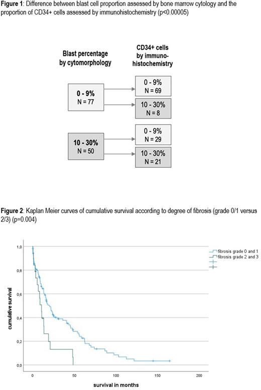
Results: The estimation of the percentage of CD34-positive cells by histopathology differed significantly from the cytomorphologically assessed medullary blast count, with a trend towards histopathological underestimation of blast cells (figure 1). Assessment of dysmegakaryopoiesis by histopathology identified 44 cases with dysplastic features, which were deemed inconspicious by cytomorphology. Histopathology yielded a significant positive correlation (p=0.002) between cellularity and degree of dysmegakaryopoiesis, and these two features were also correlated with higher-risk WHO subtypes. Fibrosis and dysmegakaryopoiesis were negatively correlated. Bone marrow cellularity seemed to be overestimated by cytomorphology compared with histopathology. The percentage of erythropoietic cells did not correlate well between cytomorphological and histopathological review. Neither WHO subtype nor medullary blast count showed an association with the proportion of erythropoiesis, regardless of the mode of evaluation. All patients with expanded erythropoiesis also showed signs of dysmegakaryopoiesis.
There was no case of cytomorphologically diagnosed MDS that was not confirmed by histopathology, and 48% of MDS diagnoses were identical as to their WHO subtype. However, in 66 cases (52%), the WHO type diagnosed by histopathology differed from the WHO type diagnosed by cytomorphology. The discrepancy was mainly due to differing estimates of medullary blast counts. In our cohort, the degree of fibrosis positively correlated with bone marrow cellularity, a finding that appeared counterintuitive to clinical experience. Increased cellularity also correlated with higher-risk MDS subgroups.
Prognosis in terms of overall survival was influenced by the following parameters: Cytomorphologically assessed medullary blast count, bone marrow cellularity, fibrosis, and percentage of erythropoiesis, the latter assessed by histopathology. Patients with of grade 2 or 3 fibrosis had an inferior outcome (figure 2).
Conclusions: In our study, histopathology was fully congruent with bone marrow cytology as far as making a diagnosis of MDS is concerned. Histopathology, though, often underestimated the blast count and showed difficulty in identifying the correct MDS type according to the WHO classification. Histopathology is also at a disadvantage regarding evaluation of single cell morphology, with the exception of assessing certain aspects of dysmegakaryopoiesis. A clear advantage of histopathology is its exclusive capability of gauging bone marrow fibrosis and cellularity, both of which are relevant for the diagnosis and prognosis of MDS. We conclude that cytomorphology and histopathology should both be harnessed to optimize the characterization of a patient's myelodysplastic syndrome.
Disclosures
Nachtkamp:Jazz Pharmaceuticals GmbH: Honoraria. Gattermann:BMS: Honoraria; Celgene: Honoraria; Takeda: Research Funding; Novartis: Honoraria. Germing:Celgene: Consultancy, Honoraria, Research Funding; Novartis: Consultancy, Honoraria.
Author notes
Asterisk with author names denotes non-ASH members.


